
Beamline 7.2W
Macromolecular Crystallography
X-ray Techniques Available at BL7.2W
BL7.2W supports multiple X-ray techniques, including:
-
Macromolecular Crystallography (MX) – Protein and biomolecule structure determination
-
X-ray Absorption Spectroscopy (XAS)
– Transmission & Fluorescence modes
-
Grazing Incidence X-ray Absorption Spectroscopy (GIXAS)
– Surface-sensitive chemical state and coordination analysis in
thin films
-
Micro-X-ray Absorption Spectroscopy (Micro-XAS)
– Local electronic and structural analysis at the micrometer scale
-
Wide-Angle X-ray Scattering (WAXS)
– Crystalline phase identification and molecular packing in bulk
and thin films
-
Grazing Incidence Wide-Angle X-ray Scattering (GIWAXS)
– Nanoscale crystallinity and orientation analysis in thin and
nanostructured films
-
X-ray Fluorescence Spectroscopy (XRF)
– Bulk and Micro mapping
-
Micro-X-ray Fluorescence (Micro-XRF)
– High-resolution elemental mapping and micro-area analysis
-
Total Reflection X-ray Fluorescence (TXRF)
– Surface and solution analysis
Each of these techniques highlights BL7.2W’s versatility, supporting research across materials science, environmental studies, nanotechnology, and life sciences.
✨ Beamline 7.2W has two main X-ray focus positions for different types of experiments. The left position is used for techniques such as MX, XAS, GIXAS, XRF, WAXS, GIWAXS, and TXRF. The right position is designed for Micro-XRF and Micro-XAS measurements.
MX, XAS, GIXAS, XRF, WAXS, GIWAXS, TXRF
2-focus position
Micro-XRF
Micro-XAS

Two X-ray focus positions at BL7.2W: the left for general techniques (MX, XAS, XRF, XRD, GIXRD, TXRF) and the right for micro-scale methods (Micro-XRF, Micro-XAS).
Detector Specifications Summary
10-cm long ion chamber (I0)
40-cm long ion chamber (I1)
40-cm long ion chamber (I2)
(All in-house development)
XAS (Transmission Mode)
-
Sample chamber isolated from ion chambers by thin polypropylene window.
-
Gas-filled in three ion chambers.
Detector
Technique
Specifications
MIC-225 Micro ion chamber
(AVS US.Inc, USA)
XAS (Transmission Mode)
-
Portable micro ionization chamber, compact for close placement to beamline components
-
Measures intensity of hard X-ray beams
-
Dimensions: 20 mm (beam direction) × 30 mm (perpendicular)
-
Housing Material: Stainless steel
-
Electrodes: Nickel-plated copper
Vortex®-ME4 Silicon Drift Detector
(Hitachi High-Tech Science America, Inc., USA)
XAS (Fluorescence Mode)
Bulk XRF
-
Four-element Silicon Drift Detector (4SDD).
-
Total active area: 200 mm².
-
Detector thickness 0.5 mm.
-
Energy resolution (FWHM): 145–260 eV at 5.9 keV.
-
Positioned at 90° relative to X-ray incident beam.
-
Accessories: Acrylic sleeves with diameters of 3, 5, 8, and 10 mm.
Vortex®-EM Silicon Drift Detector(Hitachi High-Tech Science America, Inc., USA)
Micro-XRF / Micro-XAS
-
Single-element Silicon Drift Detector (1SDD).
-
Active area: 50 mm².
-
Detector thickness 0.5 mm.
-
Energy resolution (FWHM): 124–180 eV at 5.9 keV.
-
Positioned at 45° to incident beam and sample.
-
Accessories: Acrylic sleeves with diameters of 3, 5, 8, and 10 mm.
Vortex®-90EX Silicon Drift Detector
(Hitachi High-Tech Science America, Inc., USA)
TXRF
-
Single-element Silicon Drift Detector (1SDD).
-
Active area: 50 mm².
-
Energy resolution (FWHM): 124–180 eV at 5.9 keV.
-
Motorized vertical stage with coarse and fine adjustment.
SX 165 CCD detecter
(Rayonix, L.L.C., USA)
WAXS, GIWAXS, MX
-
Equipped with MarDTB goniometer system.
-
2θ offset up to 30°
-
Active area: 61 × 61 mm².
-
Pixel size: 165 × 165 μm².
-
Readout time: 3.5 s
-
Detector-to-protein crystal distance: 45–390 mm.
-
Temperature control: 100 K using Oxford cryostream 700 series.
PILATUS3 S 1M Hybrid-photon-counting (HPC) detector
(DECTRIS Ltd., Switzerland)
WAXS, GIWAXS, MX
-
Hybrid pixel array detector.
-
Active image area: 168.7 mm × 179.4 mm.
-
Pixel size: 172 × 172 μm².
-
Readout time: 0.95 ms.
-
Detector-to-sample distance: 380–950 mm.
Absorption Edges Accessible at BL7.2W
BL7.2W supports X-ray Absorption Spectroscopy (XAS) for a wide range of elements. The chart below shows K-edge and L3-edge energies accessible at this beamline.
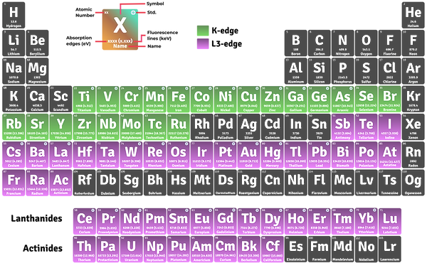
This capability enables element-specific studies such as oxidation state analysis, local structure determination, and chemical bonding investigations.
Energy Range: 4 – 22.7 keV
Photon Flux:
5 × 10¹⁰ to 1.4 × 10¹¹ ph/s/100 mA
Beam Size: 3 mm (H) × 2 mm (V)